Mitochondrial dysfunction
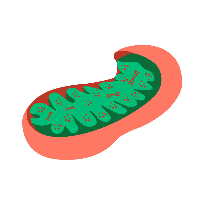
You may not know much about mitochondria, or that they play a key role in your mental health.
But these tiny organelles, which are an essential part of every one of our cells, and provide them with the energy to function via adenosine triphosphate (ATP), play a vital role in our brain and mental health.
Dysfunctional mitochondria could be contributing to mental health symptoms such as depression, anxiety, poor memory, poor concentration and attention, insomnia, and neurodegenerative diseases such as Alzheimer’s and Parkinson’s disease.
Mitochondria 101
Mitochondria are tiny organelles inside each of our trillions of cells, and they function like the batteries of each cell, producing ATP energy to power the cell’s function.
Our metabolism, hormones, central nervous system, and all our organs are powered by mitochondrial energy.
Most cells have thousands of mitochondria powering them. For example liver cells have between 400 and 4,000 and heart cells have roughly 6,000 mitochondria.
Mitochondria need two things to produce energy: food (glucose, fat or protein) and oxygen.
Interestingly, the Chinese character for Qi (vital energy) is rice (glucose) and air (oxygen). In Western science, the closest thing to the Qi of Chinese medicine, and the Prana of Ayurvedic medicine, is Mitochondrial energy.
Mitochondria evolved from bacteria, and have their own DNA, called mitochondrial DNA (mtDNA). Mitochondria provide not just more energy for their host cells, but importantly, more energy per gene.Lane, N., & Martin, W. F. (2015). Eukaryotes really are special, and mitochondria are why. Proceedings of the National Academy of Sciences of the United States of America, 112(35), E4823. Evolution of complex life requires a lot of energy to build and maintain this complexity. Mitochondria power genes to allow complex life to not just survive, but to adapt and thrive in an ever changing world.
Mitochondria have a shelf life of days for most tissue, and are designed to self-regulate, which means that when they are old, infirm or dysfunctional, they initiate mitophagy – which effectively means “mitochondrial digestion”. This is a natural process that is needed as a form of quality control to ensure mitochondria can continue to give our brain and body optimum levels of ATP.
Another part of mitochondrial quality control (or mitochondrial dynamics) is fusion and fission where mitochondria continually fuse and divide to ‘weed out’ worn out mitochondrial components which are ready for mitophagy.Chen, W., Zhao, H. and Li, Y. (2023). Mitochondrial dynamics in health and disease: mechanisms and potential targets. [online] Signal Transduction and Targeted Therapy, 8 (1), 333. Available at: https://doi.org/10.1038/s41392-023-01547-9 [accessed 21 Sep. 2026].
Mitochondria and the brain
Neurons within our brain require substantially more mitochondria than the rest of the body, due to their incredibly high energy needs.
For example, Some dopamine producing neurons have been estimated to require as many as 1 million mitochondria.Henrich, M.T., Oertel, W.H., Surmeier, D.J. and Geibl, F.F. (2023). Mitochondrial dysfunction in Parkinson’s disease – a key disease hallmark with therapeutic potential. [online] Molecular Neurodegeneration, 18 (1), 83. Available at: https://doi.org/10.1186/s13024-023-00676-7 [accessed 21 Sep. 2026].
Even though the brain makes up only 2% of our body weight, the trillions of mitochondria that the brain contains result in up to 20% of our oxygen and 25% of our total glucose being consumed by these hungry brain organelles.Faria-Pereira, A. and Morais, V.A. (2022). Synapses: The Brain’s Energy-Demanding Sites. [online] International Journal of Molecular Sciences, 23 (7), 3627. Available at: https://doi.org/10.3390/ijms23073627 [accessed 21 Sep. 2026].
Healthy mitochondria which are both abundant and functional, are necessary to produce sufficient amounts of ATP for optimal brain function and mental health.
How mitochondria can become dysfunctional
Mitochondria can become dysfunctional through excess stress, inflammation, obesity, poor diet, excessive toxins and a sedentary lifestyle.Xu, X., Pang, Y. and Fan, X. (2025). Mitochondria in oxidative stress, inflammation and aging: from mechanisms to therapeutic advances. [online] Signal Transduction and Targeted Therapy, 10 (1), 190. Available at: https://doi.org/10.1038/s41392-025-02253-4 [accessed 21 Sep. 2026].See below for more detail on root causes of mitochondrial dysfunction.
Dysfunctional mitochondria not only fail to produce enough ATP for optimal cell power, but also produce high levels of damaging ROS (Reactive Oxygen Species).
If left unchecked, excessively damaged and dysfunctional mitochondria can act as cell saboteurs and signal the whole cell to commit suicide in a process called apoptosis (programmed cell death).Dadsena, S., King, L.E. and García-Sáez, A.J. (2021). Apoptosis regulation at the mitochondria membrane level. [online] Biochimica et Biophysica Acta. Biomembranes, 1863 (12), 183716. Apoptosis is a natural process designed to protect old and dysfunctional cells, however it needs to be carefully regulated. Excessively damaged mitochondria can lead to excessive apoptosis.
How dysfunctional mitochondria affect mental health
People with depression have been shown to have impaired CNS (central nervous system) energy production, impaired neurotransmitter production, and reduced neurotrophic factors such as BDNF (brain derived neurotrophic factor) and NGF (nerve growth factor) which are essential for neural plasticity.Song, Y., Cao, H., Zuo, C., Gu, Z., Huang, Y., Miao, J., Fu, Y., Guo, Y., Jiang, Y. and Wang, F. (2023). Mitochondrial dysfunction: A fatal blow in depression. [online] Biomedicine & Pharmacotherapy, 167, 115652.
Neural plasticity can be defined as the way an individual remodels their inner neural connections according to their outer environment. Insufficient or inappropriate neural plasticity may leave an individual mentally ill-equipped to thrive in their surrounding environment.Tartt, A.N., Mariani, M.B., Hen, R., Mann, J.J. and Boldrini, M. (2022). Dysregulation of adult hippocampal neuroplasticity in major depression: pathogenesis and therapeutic implications. [online] Molecular Psychiatry, 27 (6), pp. 2689–2699. Available at: https://doi.org/10.1038/s41380-022-01520-y [accessed 21 Sep. 2026].
Impaired or dysfunctional mitochondria, dysfunctional mitophagy and excessive apoptosis are implicated in the pathology of Alzheimer’s disease and other neurological and mental health disorders.Reiss, A.B., Gulkarov, S., Jacob, B., Srivastava, A., Pinkhasov, A., Gomolin, I.H., Stecker, M.M., Wisniewski, T. and De Leon, J. (2024). Mitochondria in Alzheimer’s Disease Pathogenesis. [online] Life, 14 (2), 196. Available at: https://doi.org/10.3390/life14020196 [accessed 21 Sep. 2026].
Dysfunctional mitochondria can lead to:
- ATP is necessary for axon and neuron growth, and BDNF (Brain Derived Neurotrophic Factor) assists this growth by improving mitochondrial efficiency to increase neuronal ATPK Soman, S., Swain, M. and Dagda, R.K. (2025). BDNF-TrkB Signaling in Mitochondria: Implications for Neurodegenerative Diseases. [online] Molecular Neurobiology, 62 (2), pp. 1756–1769. Available at: https://doi.org/10.1007/s12035-024-04357-4 [accessed 21 Sep. 2026].
- when ATP production is compromised by dysfunctional or reduced numbers of mitochondria, there is decreased synaptic plasticityFaria-Pereira, A. and Morais, V.A. (2022). Synapses: The Brain’s Energy-Demanding Sites. [online] International Journal of Molecular Sciences, 23 (7), 3627. Available at: https://doi.org/10.3390/ijms23073627 [accessed 21 Sep. 2026].
- mitochondria enable neurogenesis (increased brain plasticity and regrowth) by supporting the process of maturation from stem cell to neuron, and by accumulating at the site of axon growth to provide vital ATPBüeler, H. (2021). Mitochondrial and Autophagic Regulation of Adult Neurogenesis in the Healthy and Diseased Brain. [online] International Journal of Molecular Sciences, 22 (7), 3342. Available at: https://doi.org/10.3390/ijms22073342 [accessed 21 Sep. 2026].
- NGF (Nerve Growth Factor) is required to allow for this mitochondrial accumulation
- depressed patients have been found to have either low or dysregulated levels of NGF and BDNFJaiswal, A., Shreekantiah, U. and Goyal, N. (2023). Nerve Growth Factor in Psychiatric Disorders: A Scoping Review. [online] Indian Journal of Psychological Medicine, 45 (6), pp. 555–564.
- the natural byproduct of mitochondrial energy production is ROS (Reactive Oxygen Species)
- in small amounts ROS are vital to our mental and physical wellbeing by triggering our DNA to manufacture many anti-ageing antioxidantsCheng, Y.W., Liu, J. and Finkel, T. (2023). Mitohormesis. [online] Cell Metabolism, 35 (11), pp. 1872–1886.
- excessive amounts of ROS however can undermine the enzymes and cofactors involved in the synthesis of monoamine neurotransmitters which support mental health, such as serotonin, dopamine, and noradrenalineJi, N., Lei, M., Chen, Y., Tian, S., Li, C. and Zhang, B. (2023). How Oxidative Stress Induces Depression? [online] ASN Neuro, 15, 17590914231181037.
- as we age, the activity of monoamine oxidase enzymes increases, further degrading monoamine neurotransmittersSakurai, K., Nihashi, T., Kimura, Y., Iwata, K., Ikenuma, H., Arahata, Y., Okamura, N., Yanai, K., Akagi, A., Ito, K., Kato, T. and Nakamura, A. (2022). Age-related increase of monoamine oxidase B in amyloid-negative cognitively unimpaired elderly subjects. [online] Annals of Nuclear Medicine, 36 (8), pp. 777–784. Available at: https://doi.org/10.1007/s12149-022-01760-6 [accessed 21 Sep. 2026].
- StressHassamal, S. (2023). Chronic stress, neuroinflammation, and depression: an overview of pathophysiological mechanisms and emerging anti-inflammatories. [online] Frontiers in Psychiatry, 14, 1130989., traumaLi, C. and Xiang, S. (2023). Adverse Childhood Experiences, Inflammation, and Depressive Symptoms in Late Life: A Population-Based Study. [online] The Journals of Gerontology. Series B, Psychological Sciences and Social Sciences, 78 (2), pp. 220–229.,obesityLy, M., Yu, G.Z., Mian, A., Cramer, A., Meysami, S., Merrill, D.A., Samara, A., Eisenstein, S.A., Hershey, T., Babulal, G.M., Lenze, E.J., Morris, J.C., Benzinger, T.L.S. and Raji, C.A. (2023). Neuroinflammation: A Modifiable Pathway Linking Obesity, Alzheimer’s disease, and Depression. [online] The American Journal of Geriatric Psychiatry, 31 (10), pp. 853–866., poor dietWięckowska-Gacek, A., Mietelska-Porowska, A., Wydrych, M. and Wojda, U. (2021). Western diet as a trigger of Alzheimer’s disease: From metabolic syndrome and systemic inflammation to neuroinflammation and neurodegeneration. [online] Ageing Research Reviews, 70, 101397., sedentary lifestyleWang, M., Zhang, H., Liang, J., Huang, J. and Chen, N. (2023). Exercise suppresses neuroinflammation for alleviating Alzheimer’s disease. [online] Journal of Neuroinflammation, 20 (1), 76., brain injuryObukohwo, O.M., Oreoluwa, O.A., Andrew, U.O. and Williams, U.E. (2024). Microglia-mediated neuroinflammation in traumatic brain injury: a review. [online] Molecular Biology Reports, 51 (1), 1073., gut dysbiosisO’Riordan, K.J., Moloney, G.M., Keane, L., Clarke, G. and Cryan, J.F. (2025). The gut microbiota-immune-brain axis: Therapeutic implications. [online] Cell Reports. Medicine, 6 (3), 101982., toxins and peripheral inflammationBaird, B., Noor, S., Leng, S., Ottens, A.K. and Campen, M.J. (2026). Contemporary Review: Temporal Dynamics of Neuroinflammation in Response to Environmental Exposure. [online] Toxicological Sciences2020], can all lead to CNS inflammation known as neuroinflammation
- mitochondria are both sources and targets of excess ROS during the above health challenges which can cause further neuroinflammation, leading to an increased risk of depressionSong, Y., Cao, H., Zuo, C., Gu, Z., Huang, Y., Miao, J., Fu, Y., Guo, Y., Jiang, Y. and Wang, F. (2023). Mitochondrial dysfunction: A fatal blow in depression. [online] Biomedicine & Pharmacotherapy, 167, 115652, anxietyChioino, A. and Sandi, C. (2025). The Emerging Role of Brain Mitochondria in Fear and Anxiety. [online] Current Topics in Behavioral Neurosciences, 73, pp. 33–54., insomniaAtrooz, F. and Salim, S. (2020). Sleep deprivation, oxidative stress and inflammation. [online] Advances in Protein Chemistry and Structural Biology, 119, pp. 309–336., poor memoryWięckowska-Gacek, A., Mietelska-Porowska, A., Wydrych, M. and Wojda, U. (2021). Western diet as a trigger of Alzheimer’s disease: From metabolic syndrome and systemic inflammation to neuroinflammation and neurodegeneration. [online] Ageing Research Reviews, 70, 101397., and other mental health issues
- mitochondria play a role in inflammation and growth of tissue and immune cells
- when mitochondria are in anabolic/proinflammatory mode (under threat) within microglia (resident immune cells of the Central Nervous System, or CNS) they produce ROS instead of ATPWu, Q., Tian, J., Gu, Y., Bi, X. and Zhang, H. (2026). Metabolic Reprogramming of Microglia in Neuroinflammation and Depression. [online] International Journal of Molecular Sciences, 27 (9), 3984. Available at: https://doi.org/10.3390/ijms27093984 [accessed 21 Sep. 2026].
- in this mode BDNF levels fall and undermine neural plasticity and neurogenesisWang, H., He, Y., Sun, Z., Ren, S., Liu, M., Wang, G. and Yang, J. (2022). Microglia in depression: an overview of microglia in the pathogenesis and treatment of depression. [online] Journal of Neuroinflammation, 19 (1), 132. Available at: https://doi.org/10.1186/s12974-022-02492-0 [accessed 21 Sep. 2026].
- chronic activation of microglia can lead to the death of neurons due to excessive apoptosis
- apoptosis is a mitochondria dependent processDadsena, S., King, L.E. and García-Sáez, A.J. (2021). Apoptosis regulation at the mitochondria membrane level. [online] Biochimica et Biophysica Acta. Biomembranes, 1863 (12), 183716.
What causes mitochondrial dysfunction?
In a word, “stress”, both too much and too little!
Stress is a call to the body for more energy and resources. In appropriate amounts, stress can be enlivening and leads us to be more resilient against future stresses.
Mitochondria, like the rest of our body, thrive on small amounts of challenge and stress. This is known as hormesis. Examples of hormesis are exercise, plant polyphenols, extreme heat or cold — all of which in small doses are stimulating and healthy for the mitochondria, triggering our body’s own antioxidant response. ROS generated during beneficial stress are signals for the body to boost protective measures.
When stress is excessive however, the call for energy and resources exceed the body’s psychological, biochemical and energetic reserves, leading to a decline in health and wellbeing.Liu, X., Zhang, X., Zhao, L., Long, J., Feng, Z., Su, J., Gao, F. and Liu, J. (2024). Mitochondria as a sensor, a central hub and a biological clock in psychological stress-accelerated aging. [online] Ageing Research Reviews, 93, 102145.
Mitochondria are sensitive to our environment, and, much like us, are designed to respond to states of threat or safety. Robert Naviaux coined the term “cell danger response” which is when our cells launch a biochemical chain reaction in response to stressors, sometimes continuing to do so even when the threat has passed.Naviaux, R.K. (2023). Mitochondrial and metabolic features of salugenesis and the healing cycle. [online] Mitochondrion, 70, pp. 131–163.
Peace time metabolism versus defense mode
When not presented with excessive stressors, our mitochondria are in “peace time metabolism” (a term coined by Ari Whitten of The Energy Blueprint).
Peace time metabolism
- mitochondria produce energy (ATP)
- they are in aerobic/catabolic (breakdown) mode
- they limit excessive tissue growth and reduce inflammation
However, when presented with too many stressors and energy demands, our mitochondria feel under threat and shift into “defense mode”.
Defense mode
- mitochondria stop producing energy
- they produce excessive levels of ROS (reactive oxygen species), which are oxidants
- they are in anaerobic/anabolic (build-up) mode
- this promotes tissue growth/proliferation (it can lead to the proliferation of cancerous cells for instance) and inflammation
This response is designed to protect us in the short term, however when it gets stuck on ON (as in the cell danger response) it can lead to uncontrolled cell proliferation and chronic inflammation.Naviaux, R.K. (2023). Mitochondrial and metabolic features of salugenesis and the healing cycle. [online] Mitochondrion, 70, pp. 131–163.
Unresolved chronic inflammation can lead to too much apoptosis (programmed cell death) in the case of neurons and brain health, and too little apoptosis in the case of cancer.Bertheloot, D., Latz, E. and Franklin, B.S. (2021). Necroptosis, pyroptosis and apoptosis: an intricate game of cell death. [online] Cellular & Molecular Immunology, 18 (5), pp. 1106–1121.
Ideally “defense mode” for mitochondria should be stood down as soon as a threat or demand has passed.
Excessive stress (psychological and oxidative stress) can tip mitochondria from peace time metabolism when they produce energy, to defense mode when they cause cell proliferation and inflammation.
The stressors that cause mitochondrial dysfunction
As we have seen, excessive stress can damage mitochondria. When cells get stuck in “defense mode” due to chronic and excessive stressors (be they from biochemical, psychological, or behavioural sources as listed below), it can lead to neuroinflammation in response to perceived threat.
- neurons, their blood supply and the CNS immune system all work as a ‘tight knit’ system called the “neurovascular unit”
- excessive stress can undermine the neurovascular unit and promote neuroinflammationWu, S., Yin, Y. and Du, L. (2022). Blood-Brain Barrier Dysfunction in the Pathogenesis of Major Depressive Disorder. [online] Cellular and Molecular Neurobiology, 42 (8), pp. 2571–2591. Available at: https://doi.org/10.1007/s10571-021-01153-9 [accessed 21 Sep. 2026].
- with excessive stress too many demands are made of our mitochondria and energy metabolism, exceeding our ability to produce it, which leads to high levels of ROS (reactive oxygen species) and RNS (reactive nitrogen species)
- these can damage mitochondrial DNA (mtDNA)
- small amounts of ROS are protective through mitohormesis (hormesis to the mitochondria), but large amounts can be extremely damagingCheng, Y.W., Liu, J. and Finkel, T. (2023). Mitohormesis. [online] Cell Metabolism, 35 (11), pp. 1872–1886.
- mitochondrial DNA (mtDNA) is relatively unprotected compared to cell nucleus DNA, and is easily damagedShokolenko, I. and Alexeyev, M. (2022). Mitochondrial DNA: Consensuses and Controversies. [online] DNA, 2 (2), pp. 131–148. Available at: https://doi.org/10.3390/dna2020010 [accessed 21 Sep. 2026].
- this damage to mtDNA further increases free radicals and causes ageing and degeneration, as mitochondria can’t make enough ATP for their host cells
- these dysfunctional mitochondria are usually removed through a process called mitophagy, however, high levels of mitochondrial damage can lead a cell to self-destruct (apoptosis)
- stressed mitochondria release mtDNA and cardiolipin (a type of fat only found only in mitochondria and bacteria) into the cytosol
- once outside mitochondria, these internal mitochondrial components activate the inflammasome, triggering a strong inflammatory responseXian, H., Watari, K., Sanchez-Lopez, E., Offenberger, J., Onyuru, J., Sampath, H., Ying, W., Hoffman, H.M., Shadel, G.S. and Karin, M. (2022). Oxidized DNA fragments exit mitochondria via mPTP- and VDAC-dependent channels to activate NLRP3 inflammasome and interferon signaling. [online] Immunity, 55 (8), pp. 1370–1385.e8.
mtDNA can be detected in blood plasma of depressive patients and suicide attempters, indicating that stress, mitochondrial dysfunction and neuropathology are all deeply interconnected.Park, S.S., Jeong, H. and Andreazza, A.C. (2022). Circulating cell-free mitochondrial DNA in brain health and disease: A systematic review and meta-analysis. [online] The World Journal of Biological Psychiatry, 23 (2), pp. 87–102.
Stressors to mitochondria can be biochemical, psychological, or lifestyle/behavioural:
It is the dual role of mitochondria as energy providers (catabolic mode) or suppliers of cell components (anabolic mode) which underlies a large part of how diet translates into health or disease.
The typical western diet high in calories and low in micronutrients pushes mitochondria too far into anabolic mode, turning on the glycolytic and lipogenic switches required for cell growth, proliferation and potentially, chronic inflammatory diseaseSong, M., Bai, Y. and Song, F. (2025). High-fat diet and neuroinflammation: The role of mitochondria. [online] Pharmacological Research, 212, 107615.
Overeating
- especially refined carbohydrates
- energy within food is effectively captured from sunlight via photosynthesis
- overeating can deliver more ‘sunlight energy’ than mitochondria can deal with – this is literally ‘sunburn’ on a microscopic scale!
- if mitochondria are overwhelmed with excess glucose/food, they have to export much of this extra energy and convert it into fat, and fat (or adipose tissue) in turn produces inflammatory adipokines associated with mitochondrial dysfunction and depressionHontecilla-Prieto, L., García-Domínguez, D.J., Berlanga-Gil, C., Flores-Campos, R., Muñoz-Pacheco, R., Franco-Fernández, M.D., Flores-Cordero, J.A., Sánchez-Jiménez, F., Pérez-Pérez, A., Vilariño-García, T. and Sánchez-Margalet, V. (2025). Leptin a potential link between obesity and depression. [online] Cellular and Molecular Life Sciences, 82 (1), 365. Available at: https://doi.org/10.1007/s00018-025-05892-6 [accessed 21 Sep. 2026].
Eating too often
- raises insulin and so inhibits mitochondrial quality control through mitophagy, fusion and fission
High blood sugar/glucose
- caused by excess intake of carbohydrates
- high blood sugar can be very damaging to mitochondria as it can overwhelm the mitochondrial electron transport chain, leading to damaging levels of mitochondrial ROS
- Insulin resistance, type 2 diabetes and obesity are all conditions that slow or inhibit mitophagy, meaning that individuals suffering from these conditions will often have dysfunctional, low-energy mitochondria
- samples of muscle from people with insulin resistance have been found to contain 30% less mitochondria than healthy individuals
Poor nutrition
- whole natural foods contain many of the nutrients and cofactors needed by mitochondria to metabolise food for energy
- processed ‘foods’ are usually deficient in key nutrients and antioxidants and will increase mitochondrial stress and dysfunction
Free fatty acids
- insulin resistance and obesity driven by a poor diet raise levels of free fatty acids which are associated with depression and dementiaPomytkin, I. and Pinelis, V. (2021). Brain Insulin Resistance: Focus on Insulin Receptor-Mitochondria Interactions. [online] Life, 11 (3), 262. Available at: https://doi.org/10.3390/life11030262 [accessed 21 Sep. 2026]. Song, M., Bai, Y. and Song, F. (2025). High-fat diet and neuroinflammation: The role of mitochondria. [online] Pharmacological Research, 212, 107615.
Read more about how nutritional imbalances can damage your mental health.
Exercise is a prime example, where small amounts of mitochondrial ROS (Reactive Oxygen Species) lead the body to produce its own antioxidants in response.
This mitochondrial protective response is another type of hormesis called ‘mitohormesis’.Cheng, Y.W., Liu, J. and Finkel, T. (2023). Mitohormesis. [online] Cell Metabolism, 35 (11), pp. 1872–1886.
Excess exercise
- exercise is a form of stress
- too much exercise can cause excessive ROS and inflammation, making it very difficult for mitochondria to recover and maintain their integrity
Not enough exercise
- a sedentary lifestyle deprives mitochondria of the low levels of ROS required to trigger mitohormesis, a mitochondria protective process.Memme, J.M., Erlich, A.T., Phukan, G. and Hood, D.A. (2021). Exercise and mitochondrial health. [online] The Journal of Physiology, 599 (3), pp. 803–817. Available at: https://doi.org/10.1113/JP278853 [accessed 21 Sep. 2026].
- exercise triggers the pathways necessary to build more mitochondria, in a process called mitochondrial biogenesisBurtscher, J., Millet, G.P., Place, N., Kayser, B. and Zanou, N. (2021). The Muscle-Brain Axis and Neurodegenerative Diseases: The Key Role of Mitochondria in Exercise-Induced Neuroprotection. [online] International Journal of Molecular Sciences, 22 (12), 6479. Available at: https://doi.org/10.3390/ijms22126479 [accessed 21 Sep. 2026].
- exercise can initiate protective measures that improve insulin sensitivity and help to restore mitochondrial quality control strategies, such as mitophagy, fission and fusion
Read more about the effects of inappropriate exercise on mental health.
When we sleep, our brain detoxifies largely thanks to the glymphatic system:
- neurotoxic waste in the brain is cleared by the glymphatic system during sleepChong, P.L.H., Garic, D., Shen, M.D., Lundgaard, I. and Schwichtenberg, A.J. (2022). Sleep, cerebrospinal fluid, and the glymphatic system: A systematic review. [online] Sleep Medicine Reviews, 61, 101572.
- brain mitochondria can become dysfunctional if neurotoxic waste (such as amyloid beta) accumulate due to insufficient activity of the glymphatic systemParodi-Rullán, R.M., Javadov, S. and Fossati, S. (2021). Dissecting the Crosstalk between Endothelial Mitochondrial Damage, Vascular Inflammation, and Neurodegeneration in Cerebral Amyloid Angiopathy and Alzheimer’s Disease. [online] Cells, 10 (11), 2903. Available at: https://doi.org/10.3390/cells10112903 [accessed 21 Sep. 2026].
- in an animal model of depression, glymphatic system dysfunction was found surrounding the hippocampus and cortexBarlattani, T., Grandinetti, P., Cintio, A.D., Montemagno, A., Testa, R., D’Amelio, C., Olivieri, L., Tomasetti, C., Rossi, A., Pacitti, F. and De Berardis, D. (2024). Glymphatic System and Psychiatric Disorders: A Rapid Comprehensive Scoping Review. [online] Current Neuropharmacology, 22 (12), pp. 2016–2033.
Getting to bed too late and disrupted circadian rhythms
- weakens mitochondria due to loss of mitochondrial quality control (mitophagy)Mezhnina, V., Ebeigbe, O.P., Poe, A. and Kondratov, R.V. (2022). Circadian Control of Mitochondria in Reactive Oxygen Species Homeostasis. [online] Antioxidants & Redox Signaling, 37 (10-12), pp. 647–663.
- blue light from screens is bad for mitochondria as it blocks melatoninSilvani, M.I., Werder, R. and Perret, C. (2022). The influence of blue light on sleep, performance and wellbeing in young adults: A systematic review. [online] Frontiers in Physiology, 13, 943108.
Inadequate light exposure and lack of sleep can cause hormone imbalances
- which can in turn disrupt mitochondrial functionChen, Y., Xu, W., Chen, Y., Gong, J., Wu, Y., Chen, S., He, Y., Yu, H. and Xie, L. (2024). The effect of acute sleep deprivation on cortisol level: a systematic review and meta-analysis. [online] Endocrine Journal, 71 (8), pp. 753–765.
- other hormones such as ghrelin and leptin which control appetite can also be disrupted by lack of sleep, and contribute to overeatingMosavat, M., Mirsanjari, M., Arabiat, D., Smyth, A. and Whitehead, L. (2021). The Role of Sleep Curtailment on Leptin Levels in Obesity and Diabetes Mellitus. [online] Obesity Facts, 14 (2), pp. 214–221., which as we have seen can also damage mitochondria
Inadequate light exposure and lack of sleep can cause neurotransmitter imbalance
- Gaba, serotonin, dopamine all tie in to circadian rhythmsSmith, W.K., Zhong, Z.M., Wang, W.T., Hassan, N.U., Khan, M. and Wang, H. (2025). Circadian rhythms and their roles in the pathogenesis and treatment of depression. [online] Sheng Li Xue Bao, 77 (4), pp. 689–711.
Read more about the effects of lack of nature and natural light on mental health.
- mitochondrial dysfunction in gut epithelial cells makes it very difficult for the cells to produce enough ATP to maintain the integrity of cell junctions, which can lead to leaky gutWang, Y., Lai, H., Zhang, T., Wu, J., Tang, H., Liang, X., Ren, D., Huang, J. and Li, W. (2023). Mitochondria of intestinal epithelial cells in depression: Are they at a crossroads of gut-brain communication? [online] Neuroscience and Biobehavioral Reviews, 153, 105403.
- a leaky gut can lead to inflammasome activation, and plays a role in the microbiota-inflammasome hypothesis of depressionPinzi, M., Fagiolini, A., Koukouna, D., Gualtieri, G., Rescalli, M.B., Pierini, C., Pardossi, S., Patrizio, B. and Cuomo, A. (2025). Inflammatory and Immune Biomarkers in Mood Disorders: From Mechanistic Pathways to Clinical Translation. [online] Cells, 14 (19), 1558. Available at: https://doi.org/10.3390/cells14191558 [accessed 21 Sep. 2026].
- in animal models, a cholesterol rich diet was shown to lead to cognitive decline and mitochondrial dysfunction
- high levels of cholesterol were also found in CNS mitochondrial membranes in Alzheimer’s diseaseGoicoechea, L., Conde de la Rosa, L., Torres, S., García-Ruiz, C. and Fernández-Checa, J.C. (2023). Mitochondrial cholesterol: Metabolism and impact on redox biology and disease. [online] Redox Biology, 61, 102643.
- conversely however, low plasma cholesterol has been noted in depressive patients, particularly those with lack of remission from their symptomsSong, M., Xu, P., Shang, J., Guo, Y., Cheng, Y., Lv, X., He, W., Zhai, Z. and Zhang, J. (2026). Association between total cholesterol and depression or depressive symptoms: A systematic review and meta-analysis accounting for comparator comorbidity. [online] Progress in Neuro-psychopharmacology & Biological Psychiatry, 149, 111896.
- it is difficult to understand why studies on cholesterol and neurological health are contradictory, but it could be because the brain has its own cholesterol metabolism which is largely independent from the rest of the body
- however, when the blood-brain barrier becomes ‘leaky’, unwanted peripheral cholesterol metabolites may enter the brain – and other required metabolites may become depletedHe, K., Zhao, Z., Zhang, J., Li, D., Wang, S. and Liu, Q. (2024). Cholesterol Metabolism in Neurodegenerative Diseases. [online] Antioxidants & Redox Signaling, 41 (16-18), pp. 1051–1072.
- in a similar way to leaky gut, dysfunctional mitochondria in the blood-brain barrier can make it become more leakySalmina, A.B., Kharitonova, E.V., Gorina, Y.V., Teplyashina, E.A., Malinovskaya, N.A., Khilazheva, E.D., Mosyagina, A.I., Morgun, A.V., Shuvaev, A.N., Salmin, V.V., Lopatina, O.L. and Komleva, Y.K. (2021). Blood-Brain Barrier and Neurovascular Unit In Vitro Models for Studying Mitochondria-Driven Molecular Mechanisms of Neurodegeneration. [online] International Journal of Molecular Sciences, 22 (9), 4661. Available at: https://doi.org/10.3390/ijms22094661 [accessed 21 Sep. 2026].
- dysregulated calcium levels can cause destructive excitotoxicity in neuronal cells
- loss of calcium homeostasis is a major cause of dysfunction in the mitochondria
- it can be caused by excitotoxicity (over-activation of glutamate receptors which allow excessive calcium influx), misfolded proteins and the saturated fatty acid palmitate
- mitochondria are sensitive to calcium and respond by making more ATP – excess calcium will overdrive mitochondria leading to their demise
- if calcium homeostasis is not restored, mitochondria may trigger apoptosis and destroy the whole neuron – this is excitotoxicityVerma, M., Lizama, B.N. and Chu, C.T. (2022). Excitotoxicity, calcium and mitochondria: a triad in synaptic neurodegeneration. [online] Translational Neurodegeneration, 11 (1), 3. Available at: https://doi.org/10.1186/s40035-021-00278-7 [accessed 21 Sep. 2026].
- mitochondria are both a source and target of inflammation
- when in ‘defense mode’ mitochondria shift from producing ATP to supporting pro-inflammatory cytokine productionVringer, E. and Tait, S.W.G. (2023). Mitochondria and cell death-associated inflammation. [online] Cell Death and Differentiation, 30 (2), pp. 304–312. Available at: https://doi.org/10.1038/s41418-022-01094-w [accessed 21 Sep. 2026].
- if inflammation remains unresolved, mitochondria may become dysfunctional due to mtDNA damage, compromised membranes and low activity of the electron transport chain (needed to produce ATP)Xu, X., Pang, Y. and Fan, X. (2025). Mitochondria in oxidative stress, inflammation and aging: from mechanisms to therapeutic advances. [online] Signal Transduction and Targeted Therapy, 10 (1), 190. Available at: https://doi.org/10.1038/s41392-025-02253-4 [accessed 21 Sep. 2026].
Read more about how inflammation can cause mental health issues.
Mitochondria, which have evolved from bacteria, are particularly sensitive to damage from toxins such as;
Pharmaceuticals
- statins have been shown to damage mitochondriaMollazadeh, H., Tavana, E., Fanni, G., Bo, S., Banach, M., Pirro, M., von Haehling, S., Jamialahmadi, T. and Sahebkar, A. (2021). Effects of statins on mitochondrial pathways. [online] Journal of Cachexia, Sarcopenia and Muscle, 12 (2), pp. 237–251. Available at: https://doi.org/10.1002/jcsm.12654 [accessed 21 Sep. 2026].
- antibiotics – quinolone, aminoglycoside, and β-lactam antibiotics can cause mitochondrial dysfunction
- Mitochondria originally evolved from bacteria and this common ancestry makes them particularly vulnerable to many antibioticsSuárez-Rivero, J.M., López-Pérez, J., Muela-Zarzuela, I., Pastor-Maldonado, C., Cilleros-Holgado, P., Gómez-Fernández, D., Álvarez-Córdoba, M., Munuera-Cabeza, M., Talaverón-Rey, M., Povea-Cabello, S., Suárez-Carrillo, A., Piñero-Pérez, R., Reche-López, D., Romero-Domínguez, J.M. and Sánchez-Alcázar, J.A. (2023). Neurodegeneration, Mitochondria, and Antibiotics. [online] Metabolites, 13 (3), 416. Available at: https://doi.org/10.3390/metabo13030416 [accessed 21 Sep. 2026].
Pesticides and herbicides
Pesticides and herbicides are often engineered to damage the mitochondria of pests and plants, so it’s not surprise that our own mitochondria are harmed by these chemicals.Costas-Ferreira, C., Durán, R. and Faro, L.R.F. (2022). Toxic Effects of Glyphosate on the Nervous System: A Systematic Review. [online] International Journal of Molecular Sciences, 23 (9), 4605. Available at: https://doi.org/10.3390/ijms23094605 [accessed 21 Sep. 2026].
Moulds
Mycotoxins from mould can lead to excessive levels of oxidative damage and apoptosis in mitochondria.Ehsanifar, M., Rajati, R., Gholami, A. and Reiss, J.P. (2023). Mold and Mycotoxin Exposure and Brain Disorders. [online] Journal of Integrative Neuroscience, 22 (6), 137. Available at: https://doi.org/10.31083/j.jin2206137 [accessed 21 Sep. 2026].
Infections
- VirusesJiang, T., Zhu, K., Kang, G., Wu, G., Wang, L. and Tan, Y. (2024). Infectious viruses and neurodegenerative diseases: The mitochondrial defect hypothesis. [online] Reviews in Medical Virology, 34 (4), e2565. Available at: https://doi.org/10.1002/rmv.2565 [accessed 21 Sep. 2026].
- Bacteria – mitochondria release ROS to help fight bacterial infections, but are also compromised by many bacteria.Weinberg, S.E. and Chandel, N.S. (2025). Mitochondria reactive oxygen species signaling-dependent immune responses in macrophages and T cells. [online] Immunity, 58 (8), pp. 1904–1921.
Metals
Mercury, cadmium, lead, aluminium and arsenic are all toxic to mitochondria.Sun, Q., Li, Y., Shi, L., Hussain, R., Mehmood, K., Tang, Z. and Zhang, H. (2022). Heavy metals induced mitochondrial dysfunction in animals: Molecular mechanism of toxicity. [online] Toxicology, 469, 153136.
Hydrocarbons
From smoke increase oxidative stress and mitochondrial dysfunctionAn, Z., Liu, G., Shen, L., Qi, Y., Hu, Q., Song, J., Li, J., Du, J., Bai, Y. and Wu, W. (2024). Mitochondrial dysfunction induced by ambient fine particulate matter and potential mechanisms. [online] Environmental Research, 262 (Pt 2), 119930.
Radiation
Radiation has been shown to damage mitochondria. Shimura, T., Takahashi, Y., Saito, C., Maida, R., Sasatani, M., Kunoh, T. and Ushiyama, A. (2025). Ionizing radiation triggers the release of mitochondrial DNA into the cytosol as a signal of mitochondrial damage. [online] Scientific Reports, 15 (1), 23191. Available at: https://doi.org/10.1038/s41598-025-04845-0 [accessed 21 Sep. 2026].
Endogenous metabolites
in Parkinson’s and Alzheimer’s disease misfolded proteins and neurotransmitter metabolites can be toxic to mitochondria.Choi, M.L., Chappard, A., Singh, B.P. et al. (2022). Pathological structural conversion of α-synuclein at the mitochondria induces neuronal toxicity. [online] Nature Neuroscience, 25 (9), pp. 1134–1148. Available at: https://doi.org/10.1038/s41593-022-01140-3 [accessed 21 Sep. 2026].
Psychological trauma can lead to:
Hormone and neurotransmitter imbalances
- high cortisol, whether acute or chronic, causes an increase of glucose and lipids (to fuel fight or flight) in the bloodLiu, X., Zhang, X., Zhao, L., Long, J., Feng, Z., Su, J., Gao, F. and Liu, J. (2024). Mitochondria as a sensor, a central hub and a biological clock in psychological stress-accelerated aging. [online] Ageing Research Reviews, 93, 102145.
- glucose and lipids then need to be processed by the mitochondria, resulting in metabolic stressLiu, X., Zhang, X., Zhao, L., Long, J., Feng, Z., Su, J., Gao, F. and Liu, J. (2024). Mitochondria as a sensor, a central hub and a biological clock in psychological stress-accelerated aging. [online] Ageing Research Reviews, 93, 102145.
- steroid stress hormones such as cortisol and adrenaline have a beneficial effect on mitochondria in small amounts and a detrimental effect when stress is chronic or at a high levelChoi, G.E., Lee, H.J., Chae, C.W., Cho, J.H., Jung, Y.H., Kim, J.S., Kim, S.Y., Lim, J.R. and Han, H.J. (2021). BNIP3L/NIX-mediated mitophagy protects against glucocorticoid-induced synapse defects. [online] Nature Communications, 12 (1), 487. Available at: https://doi.org/10.1038/s41467-020-20679-y [accessed 21 Sep. 2026].
- overactivation of the sympathetic nervous system during stress can undermine CNS mitochondria through loss of anti-inflammatory vagus nerve activityTan, C., Qiao, M., Ma, Y., Luo, Y., Fang, J. and Yang, Y. (2023). The efficacy and safety of transcutaneous auricular vagus nerve stimulation in the treatment of depressive disorder: A systematic review and meta-analysis of randomized controlled trials. [online] Journal of Affective Disorders, 337, pp. 37–49.
- insulin resistance
- chronically high glucose levels due to chronically high stress can lead to insulin resistanceSharma, K., Akre, S., Chakole, S. and Wanjari, M.B. (2022). Stress-Induced Diabetes: A Review. [online] Cureus, 14 (9), e29142. Available at: https://doi.org/10.7759/cureus.29142 [accessed 21 Sep. 2026].
- low thyroid hormones – T3 and T2
- T3 and T2 support oxidative phosphorylation (the oxidation of nutrients to make ATP)
- T3 supports mitochondrial biogenesis
- low estrogen
- estrogen, acting through estrogen receptors, supports oxidative phosphorylation, mitochondrial biogenesis, fission and fusionKlinge, C.M. (2020). Estrogenic control of mitochondrial function. [online] Redox Biology, 31, 101435. Available at: https://doi.org/10.1016/j.redox.2020.101435 [accessed 21 Sep. 2026].
Increased production of ROS
- chronic psychological stress drives the production of ROS in mitochondria, by undermining oxidative phosphorylation (the oxidation of nutrients to produce ATP) in mitochondriaLiu, X., Zhang, X., Zhao, L., Long, J., Feng, Z., Su, J., Gao, F. and Liu, J. (2024). Mitochondria as a sensor, a central hub and a biological clock in psychological stress-accelerated aging. [online] Ageing Research Reviews, 93, 102145. Available at: https://doi.org/10.1016/j.arr.2023.102145 [accessed 21 Sep. 2026].
Mitochondrial DNA (mtDNA) leaking into the bloodstream
- people exposed to psychological stress have been shown to have mtDNA in their bloodPark, S.S., Jeong, H. and Andreazza, A.C. (2022). Circulating cell-free mitochondrial DNA in brain health and disease: A systematic review and meta-analysis. [online] The World Journal of Biological Psychiatry, 23 (2), pp. 87–102. Available at: https://doi.org/10.1080/15622975.2021.1938214 [accessed 21 Sep. 2026].
- mtDNA can be detected in blood plasma of depressive patients and suicide attempters
- components of mitochondria, such as mtDNA, are seen as danger signals once outside mitochondria – don’t forget that mitochondria used to be bacteria
- escaped mitochondrial components are known as DAMPs (damage-associated molecular pattern) and trigger an inflammatory immune responseNewman, L.E. and Shadel, G.S. (2023). Mitochondrial DNA Release in Innate Immune Signaling. [online] Annual Review of Biochemistry, 92, pp. 299–332. Available at: https://doi.org/10.1146/annurev-biochem-032620-104401 [accessed 21 Sep. 2026].
- ATP inside a cell provides the cell with energy, but once outside a cell, along with its products ADP, AMP and adenosine, it forms part of the purinergic signalling system
- imbalances in the purinergic system are seen in depression and, interestingly, it is thought that caffeine exerts its antidepressant effects by blocking purinergic receptorsZhao, Y.F., Verkhratsky, A., Tang, Y. and Illes, P. (2022). Astrocytes and major depression: The purinergic avenue. [online] Neuropharmacology, 220, 109252. Available at: https://doi.org/10.1016/j.neuropharm.2022.109252 [accessed 21 Sep. 2026].
Read more about the effects of psychological trauma on mental health.
As we age we lose mitochondrial function
- ageing results in increased mtDNA mutations and reduced mitochondrial ATP productionXu, X., Pang, Y. and Fan, X. (2025). Mitochondria in oxidative stress, inflammation and aging: from mechanisms to therapeutic advances. [online] Signal Transduction and Targeted Therapy, 10 (1), 190. Available at: https://doi.org/10.1038/s41392-025-02253-4 [accessed 21 Sep. 2026].
- it is estimated that age-related neuronal reductions in ATP that exceed 20% may lead to neurological disease
- the good news is that exercise can initiate mechanisms to repair mutated mtDNA and stimulate mitochondrial biogenesis
- appropriate exercise is therefore essential throughout life, particularly in our later years, to keep our neurons and synapses energised and free from diseaseMemme, J.M., Erlich, A.T., Phukan, G. and Hood, D.A. (2021). Exercise and mitochondrial health. [online] The Journal of Physiology, 599 (3), pp. 803–817. Available at: https://doi.org/10.1113/JP278853 [accessed 21 Sep. 2026].